
Case Study
Company |
Industry |
 |
Healthcare |

Our client, a medical startup based in the US, is dedicated to extracting insights from different types of medical images. With the help of AI, their objective is to build a product that can automatically determine what each pixel represents in the medical images. This AI solution has the potential to significantly improve the efficiency and precision of medical professionals' diagnostic processes.
We have developed multiple neural network models to accurately segment different human organs on medical images. This solution has substantially accelerated the diagnostic capabilities of professional radiologists, enabling them to promptly identify medical abnormalities. All data used for model development and evaluation was fully anonymized to ensure patient privacy and compliance with ethical standards.


The project involved the analysis of various types of medical images. These images included CT scans (Computerized Tomography) and MRIs (Magnetic Resonance Imaging). The primary goal was to develop neural network models that automatically distinguish one human organ from another.
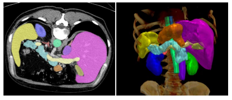 Human organ segmentation in 2D and 3D (CT)
nnU-Net: a self-configuring method for deep learning-based biomedical image segmentation
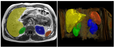 Human organ segmentation in 2D and 3D (MRI)
nnU-Net: a self-configuring method for deep learning-based biomedical image segmentation
The main problem with medical image segmentation is that the model should determine for each pixel on the image what is the corresponding organ. This means determining whether a pixel belongs to the liver, pancreas, or constitutes part of the background.
To facilitate this, the client had already conducted labeling on select images, with the assistance of expert radiologists and other trained annotators. The idea of the project was to create an AI model capable of replicating these human annotations, a process that demands considerable time and labor resources. Most importantly, the AI model needed to perform effectively on entirely new, unseen images, which were not part of the model-building dataset.
A major challenge in medical segmentation projects is transitioning from a model that performs well on familiar images to unseen images. Improving from moderately accurate to significantly high accuracy requires expert AI model development skills.
Medical image segmentation is complicated by its inherently three-dimensional nature. It involves multiple sequential image scans that describe a human organ in 3D, making individual 2D image scans not independent.
Images originate from diverse medical machines made by different manufacturers, each using varying scanning protocols. Additionally, the images come in different modalities (MRI and CT), with varying levels of annotation quality and availability for different organs.
Assessing and adapting approaches from various scientific papers poses a continuous challenge. The AI research area is constantly expanding, offering an exponentially increasing array of options for achieving high accuracy. However, each experiment requires substantial GPU computing time, requiring careful planning to balance choices with their associated costs.


A complete data science and machine learning pipeline was developed to generate multiple optimized neural network models for image segmentation. The pipeline automatically prepares data, runs experiments, and evaluates results, leading to the creation of better models through collaboration with the client to enhance training data. The models operate on individual or sequences of images, or both, to input unlabeled images and generate segmentation masks for each pixel. The predicted masks were in great agreement with the ground truth expert annotations, fulfilling the project's main goal.
 Python
 PyTorch
 MONAI
MONAI
 Albumentations
 Numpy
Numpy
 OpenCV
 Tensorboard
 ITK-Snap

The models achieved 97% accuracy in reproducing human annotations using the standard Dice coefficient in both 2D and 3D images. Additionally, the models had a 96% agreement with expert annotations in predicting organ volumes, enhancing the efficiency of diagnosing medical abnormalities. The client successfully demonstrated the usefulness of the model predictions to radiologists, improving their diagnostic capabilities.
Discover the impact of our custom AI solutions on business success through customer stories
